The Head Shape Evaluation Clinic is a unique collection of multidisciplinary clinical experts dedicated to the evaluation and treatment of problems leading to abnormal head shape in the infant and related causes. These abnormal shapes are divided into synostotic(rare) and nonsynostotic causes.
Synostosis implies that there has been an abnormal fusion of some of the cranial bones leading to abnormal growth and shape. This is a rare event and is typically thought to occur in approximately one in 3000 live births. These conditions often require a surgical intervention to correct but are fortunately seldom in occurrence.
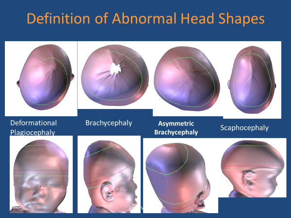
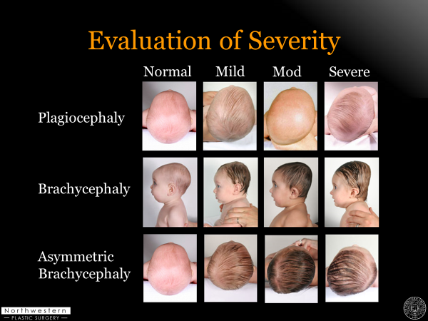
The most common cause of abnormal head shape is “positional molding” and the two common presentations of positional molding are Plagiocephaly and Brachycephaly. Plagiocephaly means “flat head” and is a right or left sided problem. Brachycephaly means “short head” which presents as a flattening in the back of the head. These asymmetries can present together and are then referred to as an asymmetric brachycephaly. 
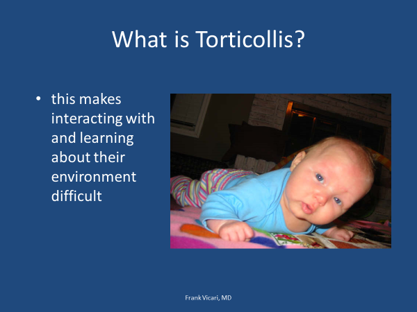
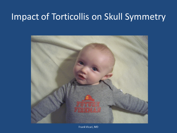
These problems are commonly associated with set of muscle weaknesses referred to as Torticollis.
Background:
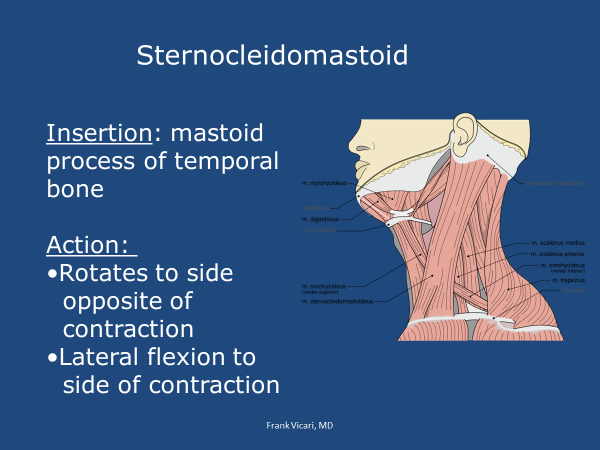
The shape of our head is determined by the forces acting on it. These forces are the push of the brain from the inside (brain growth) and the pull of the various muscle groups acting on the outside. The muscles that turn the head from left to right are called the Sternocleidomastoid muscles (SCM) and attach from behind the ear and to the clavicle and sternum in front. If these muscles are not acting equally and together (Torticollis), the infant will have difficulty turning his head to one side and will end up having a sleeping preference to the opposite side. You can also see a tilt of the head to one side. If you look at the child from the back, you may notice one ear is closer to the shoulder than the other. This can worsen the facial asymmetry noted with plagiocephaly.
The muscles that extend the neck begin in the back and attach approximately halfway up the back of the head. If they are week there tends to be a flat spot directly in the back of the head.
The importance of having these large muscle strong and working in coordination is why our first line of defence for positional cranial molding is to strengthen these muscles and why we feel Physical Therapy and a consistent home program, demonstrating how to encourage tummy time, is so important to correcting and avoiding these problems.
Your Clinic Experience:
When you visit our multidisciplinary clinic, you will be seen by a craniofacial surgeon, an experienced physician assistant, and a physical therapist or occupational therapist trained in the diagnosis and management of torticollis. You will also be evaluated by state-of-the-art equipment which will provide quick and accurate imaging of your child’s head shape. At the end of your appointment you will have a diagnosis and treatment plan.
The plan will be customized for your child and may include a home program, a recommendation for physical therapy or occupational therapy and may also include a recommendation for an orthotic cranial molding helmet. We typically recommend an orthotic from Orthomerica (www.orthomerica.com).
A “helmet” is recommended for less than 1/3rd of our patients, which means that approximately 70% of the time we can manage the problem with physical therapy and a home program alone. This typically depends upon the severity of the asymmetry at the age at which the child is seen in our clinic.
Telemedicine appointments are also available upon request.